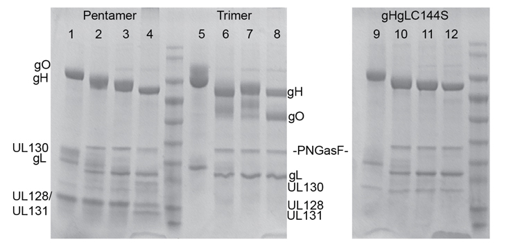
Supplemental Figure one: Deglycosylation of gH/gL protein complexes. Lanes 1, 5, and 9 are pentamer, trimer and gHgLC114S without PNGase F treatment. Lanes 2, 6, and 10 are gHgL protein complexes after 1h PNGase F treatment. Lanes 3, 7 and 11 are gHgL protein complexes after 3h PNGase F treatment. Lanes 4, 8 and 12 are gHgL proteins complexes pre-denatured before PNGase F treatment, and then incubated with PNGase F overnight. PNGase F treatment was performed according to the manufacturer’s instructions (NEB).
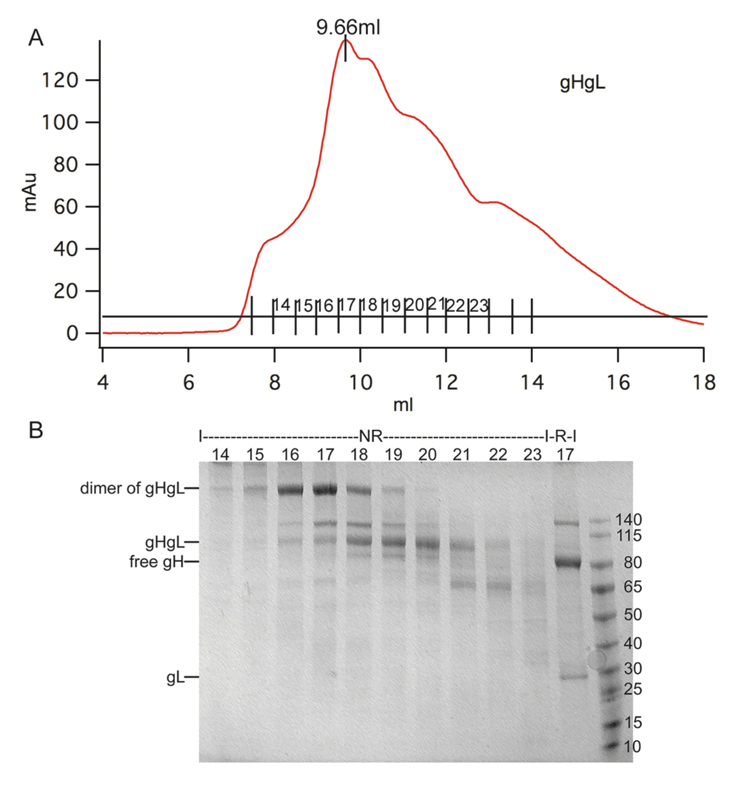
Supplemental Figure two: (A) Gel filtration analysis of a purified prep of wtgHgL (A prep consists of both gHgL dimers as well as gH/gL monomers). (B) SDS-PAGE analysis of each fraction from the gel filtration elution. NR: non-reducing SDS-PAGE loading buffer (NO TCEP); R: reducing SDS-PAGE loading buffer (with TCEP). Based on the SDS-PAGE gel analysis, we collected fractions 15- 17, and fractions 18-20 separately for further studies.