Colon Cancer Stages
Doctors label cancer by stages. Stages show how much cancer is in your body, how far it has spread and how quickly it may grow.
Stages help doctors and patients understand how serious cancer is. It also helps your Knight Cancer Institute care team know how to best treat it.
What is cancer staging?
With colon cancer, stages show:
- Where the cancer is in the colon
- Whether it has broken through the wall of the colon
- If it has spread to your lymph nodes
- If it has spread to other organs like the lungs or liver
Doctors determine colon cancer stages through:
- Physical exam
- Imaging tests, such as an MRI scan (magnetic resonance imaging)
- Biopsies (tissue samples looked at under a microscope)
Understanding colon cancer stages
The lower the stage number, the less the cancer has spread. For example:
- A stage 0 cancer is the earliest stage. Abnormal cells are in only the colon’s first layer.
- A stage IV, or 4, is advanced colon cancer. It has spread to a distant area, organ or lymph node.
Staging cancer is complicated. You can ask your care team to explain it in ways that you understand.
Stage 0
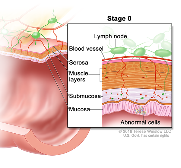
Abnormal cells are in the colon's lining, the mucosa.
Stage I
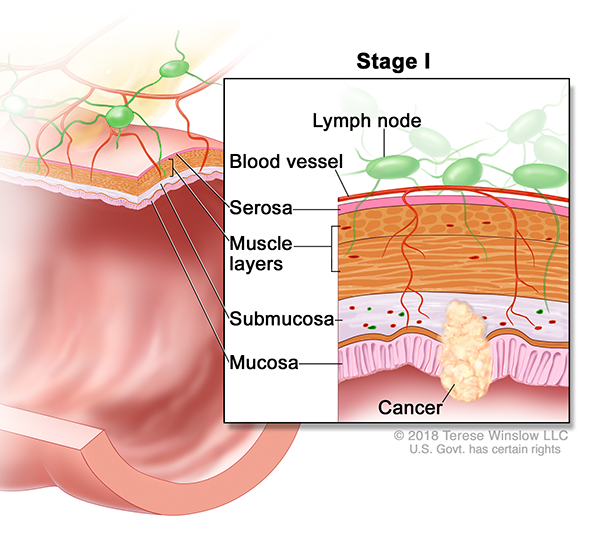
The tumor has grown into the second layer of the colon, the submucosa, or to the third layer, the muscle layer.
Stage II
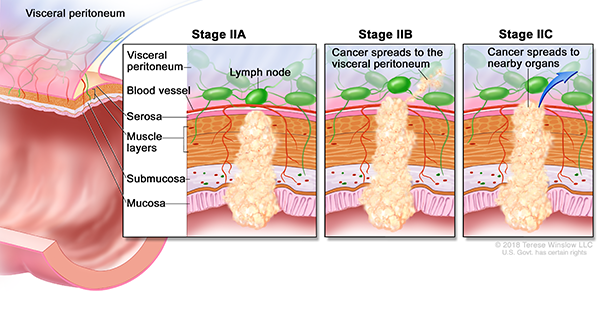
Stage IIA: The tumor has reached the colon’s outer layer, the serosa.
Stage IIB: The tumor has reached the tissue that surrounds organs in the belly, the visceral peritoneum.
Stage IIC: The tumor has grown through the colon to nearby organs.
Stage III
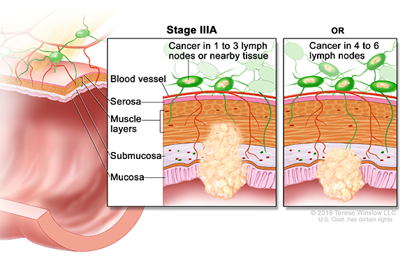
Stage IIIA: One of these:
- The tumor has grown into the submucosa and possibly the muscle layer. Cancer is also in one to three nearby lymph nodes, or cancer cells have clustered near lymph nodes.
- The tumor has grown to the submucosa, and cancer is in four to six nearby lymph nodes.
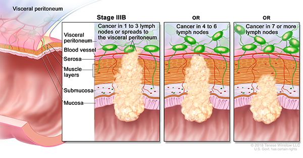
Stage IIIB: One of these:
- The tumor has spread to the serosa or to the visceral peritoneum. Cancer is also in one to three nearby lymph nodes, or cancer cells have clustered near the lymph nodes.
- The tumor has spread to the muscle layer or to the serosa, and cancer is in four to six nearby lymph nodes.
- The tumor has grown to the submucosa or to the muscle layer, and cancer is in seven or more nearby lymph nodes.
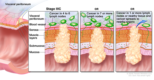
Stage IIIC: One of these:
- The tumor has spread to the visceral peritoneum, and cancer is in four to six nearby lymph nodes.
- The tumor has spread into the serosa or reached the visceral peritoneum, and cancer is in seven or more nearby lymph nodes.
- The tumor has spread to nearby organs. Cancer is also in one or more nearby lymph nodes, or cancer cells have clustered near the lymph nodes.
Stage IV
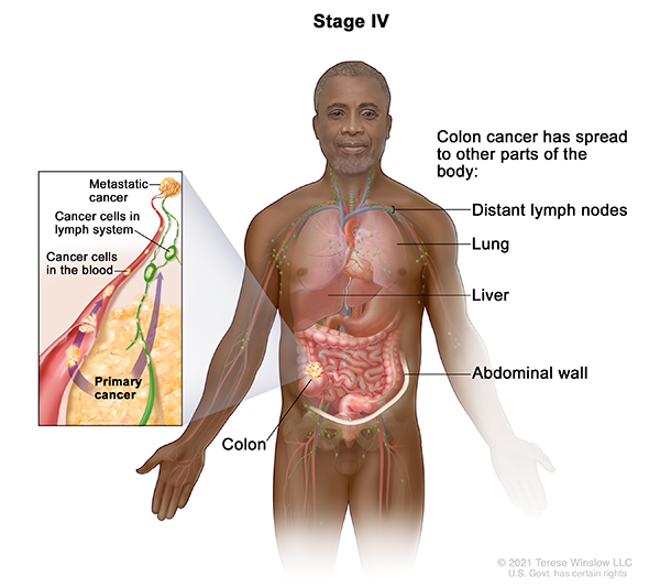
Stage IVA: Cancer has spread to one distant area, organ or set of lymph nodes.
Stage IVB: Cancer has spread to more than one distant area, organ or set of lymph nodes.
Stage IVC: Cancer has spread to distant parts of the belly lining (the peritoneum) and may have spread to other distant areas, organs or lymph nodes.
For patients
Call 503-494-7999 to:
- Request an appointment
- Seek a second opinion
- Ask questions
Location
Knight Cancer Institute, South Waterfront
Center for Health & Healing, Building 2
3485 S. Bond Ave.
Portland, OR 97239
Free parking for patients and visitors
Refer a patient
- Refer your patient to OHSU.
- Call 503-494-4567 to seek provider-to-provider advice.
Cancer clinical trials
Clinical trials allow patients to try a new test or treatment.
Read more
Learn more about OHSU Knight Cancer Institute treatments: