MR Epilepsy/Seizure WWO Neuro Protocol
Last updated:3/21/2019
Charge as: Brain WWO
Scanner preference: 3T only
Coil: Head
| Plane | Weighting | Mode | Slice | Gap | FAT SAT | FOV | Notes |
|---|---|---|---|---|---|---|---|
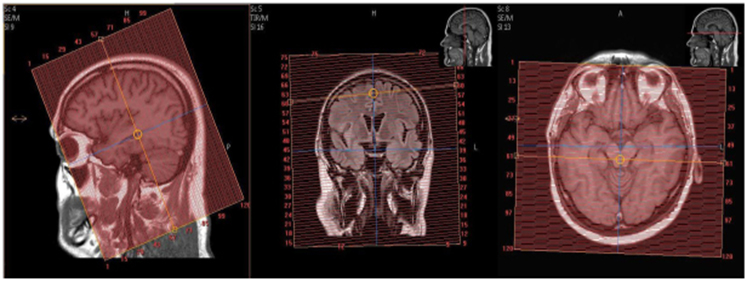
| AXIAL | T2* | GRE | 3mm | 1mm | None | 23cm | Parallel to Temporal Lobe |
| AXIAL | T2 | TSE | 3mm | 1mm | None | 23cm | Parallel to Temporal Lobe |
| AXIAL | DWI 2mm Voxel | SE EPI | 3mm | 0.3mm | SPIR | 23cm | Angle to Corpus- Skull Base to Vertex |
| OBL COR | T2 STIR | TSE | 2mm | 0.2mm | STIR | 23cm | Whole brain, perpendicular to temporal lobe |
| OBL COR | FLAIR | TSE | 3mm | 1mm | None | 23cm | Whole brain, perpendicular to temporal lobe |
| SAG | 3D T1 MPRAGE | 3D TFE | 1mm | 1mm | None | 23cm | Extend slice coverage 1-2 mm beyond the skin margin, not necessarily to cover the ears. This ensures adequate coverage/signal on the AX and COR MPRs. Generate OBL COR and AXIAL MPRs from this sequence. |
| AXIAL MPR | MPR | 3D TFE | 1.3mm | 1.3mm | None | 23cm | Angle to Corpus. Reconstruct as 1.3mm skip 1.3mm. |
| OBL COR MPR | MPR | 3D TFE | 1mm | 1mm | None | 23cm | Whole brain, perpendicular to temporal lobe |
| Optional: AX | (GPI) MPRAGE | 3D | ISO | ISO | None | Do not change | Optional if requested: DO NOT ANGLE. Limited Coverage. Cover inferior aspect of temporal lobes to vertex. Scan time will be long (6-8 Minutes). Run on 3T scanners only. |
Contrast injection
| Plane | Weighting | Mode | Slice | Gap | FAT SAT | FOV | Notes |
|---|---|---|---|---|---|---|---|
| AXIAL | T1 | TSE | 4mm | 1mm | None | 23cm | Angle to Corpus - Skull Base to Vertex |
| COR | T1 FAT SAT | TSE | 4mm | 1mm | SPIR | 23cm | Cover frontal through Occipital Bone |